
 Türkçe
 Deutsch
 Français
“Eye treatments” refers to all medical interventions used to protect and improve visual function or to manage diseases that impair eye health. This covers a wide range of conditions, from clarity problems in the eye’s optical system to pressure imbalances in the eye’s internal structure or damage to the nerve layer. The main purpose of treatments is to optimize an individual’s ability to see, provide clear and healthy vision, and improve quality of life.
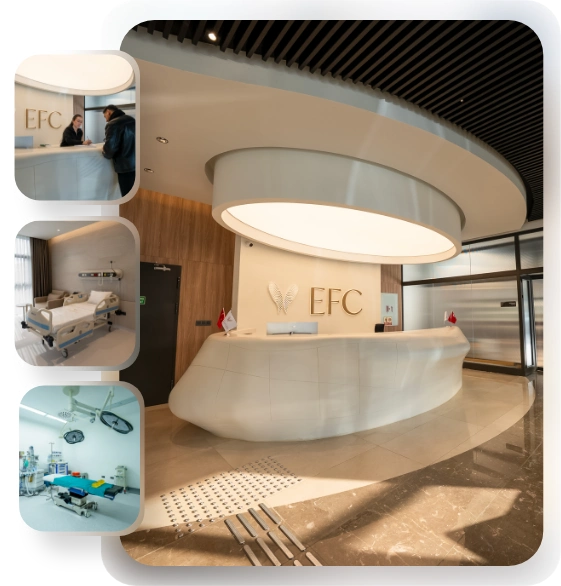
EFC CLINIC
Comprehensive Care: From Initial Consultation to Follow-Up.
EFC CLINIC is a center of excellence specializing in the most meticulous fields of surgical medicine, from aesthetic surgery to interventional treatments—where every step progresses with refined attention. Medical excellence, aesthetic precision, and uncompromising ethical standards converge on the same path. Our subspecialty-trained experts aim to achieve natural and reliable results by delivering evidence-based care supported by modern imaging, standardized protocols, and safety systems. From consultation to recovery, your care is coordinated end-to-end with clear communication, transparent planning, and genuine respect for your health.
Which laser methods are used to get rid of glasses or contact lenses?
Glasses-free surgeries, namely laser vision correction, are procedures that permanently change the shape of the cornea—the outermost transparent layer of the eye—to correct refractive errors such as myopia (distance), hyperopia (near), and astigmatism. Today, three main modern methods stand out.
- PRK / Trans-PRK (No-Touch)
- LASIK (Flap Method)
- SMILE (Flapless Method)
PRK / Trans-PRK (No-Touch) is the oldest and biomechanically (corneal strength) most reliable laser technique. In this method, the epithelium layer at the very top of the cornea, which can regenerate itself, is removed and the laser is applied directly to the underlying tissue. In the “Trans-PRK” (No-Touch) version, this removal is also performed with a laser (contact-free). Since no flap is created in the cornea, corneal strength is preserved at the highest level. However, the healing time is longer than the other methods, and the first few days after surgery may be more painful or stinging.
LASIK (or its modern version Femto-LASIK) is the most widely performed method in the world. In this procedure, a thin flap is created on the upper part of the cornea with a “femtosecond” laser. This flap is opened to the side like a door, the laser is applied to the underlying tissue, and the flap is closed back into place. Its biggest advantage is that healing is very fast; patients usually start to see clearly the next day. Its disadvantages are that this flap can create a lifelong sensitivity to impacts (even if with a low probability) and, due to cutting of the corneal nerves, it can cause more dry eye than the other methods.
SMILE is the newest laser technology. In this method, no flap is created in the cornea. The femtosecond laser creates a small disc-shaped tissue (lenticule) inside the cornea, under an intact layer of tissue, according to the amount of correction. The surgeon then removes this piece of tissue by pulling it out through a very small 2–4 mm incision made at the edge of the cornea. Thanks to the absence of a flap, there are no LASIK-related risks and the risk of dry eye is lower. Visual recovery is slightly slower than LASIK, but significantly faster than PRK.
The choice of method depends on the patient’s corneal thickness, prescription, dry eye status, and lifestyle (for example, whether they do active sports).
Does having ‘Xtra’ (Cross-Linking) together with laser treatment increase protection?
“Laser Xtra” (for example LASIK Xtra) is the application of accelerated corneal cross-linking (CXL) to the cornea in the same session during laser vision correction. The theoretical aim of this procedure is to strengthen the corneal tissue, which is thinned to some extent by the laser, and to reduce to zero the risk of a serious complication called “keratectasia” (forward bulging of the cornea) in the future.
However, scientific research on the effectiveness and safety of this protective approach is quite conflicting. While it is expected to strengthen the cornea, some large-scale analyses in recent years have shown that instead of providing benefit, this procedure may further reduce corneal thickness or negatively affect long-term visual outcomes. These findings indicate that “Xtra” protocols applied as standard together with laser are not yet ideal. Therefore, CXL-Xtra is not a procedure routinely recommended for every patient, and its benefit has not yet been clearly proven.
If I am not suitable for laser, is there a way to correct high prescriptions?
Yes, there is. Especially for patients whose corneas are too thin to allow laser, or whose myopia/hyperopia values are far above the correction limits of laser (for example, myopia higher than 8–10 diopters), “Phakic Intraocular Lenses” (PIOLs) are an excellent alternative.
These lenses are also called “Implantable Collamer Lenses” (ICL), popularly known as “permanent intraocular lenses.” They are different from the lenses used in cataract surgery; the patient’s own natural, transparent lens is not touched. PIOLs are high-tech, soft, biocompatible lenses placed just in front of the patient’s own lens.
In this method, no corneal tissue is removed (as with laser); on the contrary, the defect is corrected by adding (additive) to the eye. Its reliability and effectiveness are very high. Reliable scientific comparisons performed for high myopia patients have shown that PIOL (ICL) surgery is safer than laser. In addition, PIOLs provide better “quality of vision” (especially night vision and contrast sensitivity) compared to laser.
These lenses can even be used as an option to correct refractive error in patients with corneal disease (keratoconus) who are not suitable for laser. The main risk of this method is that (with a low probability) it may trigger early cataract development due to its proximity to the patient’s own natural lens. However, modern lens designs have minimized this risk.
Contact us now to get detailed information about our treatments and procedures and to schedule an appointment!
How can we stop the progression of keratoconus?
Keratoconus is the progressive thinning and forward protrusion of the cornea that impairs vision. This condition results from weakening of the cornea’s biomechanical structure. Today, the only proven treatment method that can stop the progression of this disease is “Corneal Cross-Linking” (CXL).
This treatment is a procedure aimed at strengthening the corneal structure. Vitamin B2 (Riboflavin) drops are instilled into the eye, and then ultraviolet-A (UV-A) light at a specific wavelength is applied. The interaction of these two agents enables the formation of new chemical bonds (“cross-links”) between collagen fibers in the cornea. These new bonds increase the mechanical resistance and stiffness of the cornea, preventing disease progression and further protrusion of the cornea.
There are different application protocols (methods) for the treatment.
- Conventional Method (Epi-off / Debridement)
- Accelerated Method (Accelerated CXL)
- Transepithelial Method (Epi-on / Transepithelial)
The “Epi-off” (debridement) method is the original method, accepted as the “gold standard,” performed by removing the epithelium, which is the uppermost layer of the cornea. It allows riboflavin to penetrate the tissue in the best way and creates the highest biomechanical effect. This is the method with the strongest results, but since the epithelium is removed, the first few days after surgery are painful and the healing time is longer.
“Epi-on” methods are performed while preserving the epithelium. In this way, there is no post-operative pain, healing is very fast, and the risk of infection is minimal. However, since the epithelium acts as a barrier, it is harder for riboflavin to pass through, and it is accepted that its effectiveness may be somewhat lower compared to the de-epithelialized method.
Which method is chosen is determined according to the severity of the disease and the patient’s comfort expectations.
What is corneal transplantation (keratoplasty) and what types are there?
Corneal transplantation (keratoplasty) is a surgical procedure in which diseased or damaged corneal tissue is replaced with healthy donor corneal tissue. In the last twenty years, corneal transplant surgery has undergone a radical change. The “full-thickness” (PKP) transplants that used to be standard have now been replaced by “lamellar” (layered) transplants, in which only the diseased layer is replaced.
Corneal transplant techniques are as follows:
- PKP (Penetrating Keratoplasty / Full-Thickness Transplant)
- DALK (Deep Anterior Lamellar Keratoplasty / Anterior Layer Transplant)
- EK (Endothelial Keratoplasty / Posterior Layer Transplant)
PKP (Full-Thickness Transplant) is the traditional method. All layers of the cornea (full thickness) are removed and the donor cornea is sutured in place.
DALK (Anterior Layer Transplant) is preferred especially in conditions where the anterior and middle layers of the cornea (stroma) are diseased, such as keratoconus, but the innermost endothelial layer is healthy. In this technique, the patient’s own healthy endothelial layer is preserved. This is a major advantage, because since the patient’s own endothelium is preserved, there is no long-term cell loss and the risk of graft rejection is lower.
EK (Posterior Layer Transplant) is the most frequently performed type of transplant today. It is used to replace only the diseased innermost layer of the cornea (endothelium). It is the standard treatment for endothelial failure conditions such as Fuchs Dystrophy or corneal edema that can develop after cataract surgery. It also has types such as DSAEK and DMEK:
When these two posterior layer transplants are compared, DMEK stands out by far as the best method. In DMEK, only the endothelium and its membrane (very thin tissue) are transplanted. Compared to DSAEK, visual acuity is much better and recovery is much faster. Its biggest advantage is that the risk of graft rejection is 15 to 20 times lower than both DSAEK and full-thickness (PKP) transplantation. The only disadvantage is that since the transplanted membrane is very thin, the risk of post-operative detachment (decollement) is somewhat higher; however, this can usually be easily corrected in an office setting by injecting air into the eye again.
Cataract and Lens Surgery
Is there a difference between the ‘laser method’ (FLACS) and the ‘Phaco’ (PCS) method in cataract surgery?
Cataract is the loss of transparency and clouding of the eye’s natural lens. Its treatment is surgical removal of this cloudy lens and implantation of an artificial intraocular lens (IOL). Today, two main modern techniques are used for this procedure: Traditional “Phacoemulsification” (PCS) and “Femtosecond Laser-Assisted Cataract Surgery” (FLACS).
Traditional Phaco (PCS) is the sutureless method that has been the “gold standard” for years. The surgeon uses a “Phaco” device that employs ultrasound (sound wave) energy to break up and aspirate the cloudy lens inside the eye.
The Laser Method (FLACS), on the other hand, is a technique in which some critical steps of the surgery (corneal incisions, opening of the anterior capsule of the lens, and fragmentation of the lens) are performed automatically by a computer-controlled femtosecond laser instead of the surgeon’s hand. After the laser completes these steps, the surgeon still uses the Phaco device to aspirate the fragmented lens.
So, is this expensive laser technology (FLACS) better than traditional Phaco? Large, multicenter, high-quality scientific studies conducted to answer this question have directly compared the two methods. The results are quite clear: No significant difference has been found between the two methods in terms of the patient’s final visual success. Although the laser method (FLACS) automates some steps of the surgery, it has been shown that it does not provide an extra contribution to the patient’s visual acuity or the speed of recovery (in the long term).
In modern cataract surgery, it is understood that the main factor determining refractive success (clear vision without glasses) is not how the lens is removed (Laser or Phaco), but which lens technology is implanted.
Which intraocular lens (IOL) technologies are available in cataract surgery?
The most important factor determining success in cataract surgery is the selection of the artificial lens (IOL) implanted in the eye. This choice directly determines the patient’s need to use glasses after surgery.
Lens technologies are ranked according to the patient’s expectation of becoming free from glasses.
- Monofocal (Single-Focus) Lenses
- Toric (Astigmatism-Correcting) Lenses
- EDOF (Extended Depth of Focus) Lenses
- Trifocal (Three-Focus / “Smart”) Lenses
Monofocal (Single-Focus) Lenses are standard, traditional lenses. They provide only one focal point (usually distance). Patients see distant vision clearly without glasses after surgery, but they must use glasses for intermediate (computer) and near (reading) distances.
Toric (Astigmatism-Correcting) Lenses are essential for patients who have high and regular astigmatism in the cornea. The lens has built-in astigmatism correction power. If a toric lens is not implanted in an astigmatic patient, the patient will still need to wear astigmatic glasses to see distance clearly after surgery.
EDOF (Extended Depth of Focus) Lenses work by “stretching” a single focal point rather than splitting light into multiple points. In this way, they provide continuous and high-quality vision from distance to intermediate range (computer, kitchen counter).
Trifocal (Three-Focus) Lenses, popularly known as “smart lenses,” split light into three focal points—distance, intermediate, and near. They offer patients the highest level of independence from glasses.
Contact us now to get detailed information about our treatments and procedures and to schedule an appointment!
What are the disadvantages of ‘smart lenses’ (EDOF and Trifocal)?
These advanced technology lenses (EDOF and Trifocal) that correct presbyopia (age-related near vision loss) come with a “cost” for the glasses-free life they provide. This cost is unwanted light perceptions called “photic phenomena.”
Because these lenses split or extend light to different focal points, they can create halos, glare, or starbursts around light sources such as headlights and street lamps, especially at night.
Trifocal (three-focus) lenses generally have a higher frequency of these photic phenomena because they provide three focal points. In return, they offer the patient the highest level of independence from glasses (including near reading).
EDOF lenses, on the other hand, are designed to reduce these side effects and are generally considered to cause less halos and glare than trifocal lenses. They offer high-quality distance and intermediate (computer) vision, but a reading glass may be needed to read very fine print (e.g., a medication leaflet).
This is a matter of “preference,” depending on lifestyle, that should be discussed in detail between the patient and the doctor.
Glaucoma (Intraocular Pressure) Management
What is the first step in the treatment of intraocular pressure (glaucoma)?
Glaucoma (intraocular pressure) is a chronic disease that causes progressive and irreversible damage to the optic nerve. The goal of treatment is to stop or slow the progression of nerve damage by lowering intraocular pressure (IOP).
The first step is to determine an individual “Target Intraocular Pressure” for each patient. This target is not a fixed number; it is determined according to the patient’s condition. The more advanced the disease is, the higher the baseline pressure is, or the younger the patient is, the lower the target pressure is set.
To reach this target, there are two “first-line” (initial preference) treatment options: eye drops or SLT laser. In open-angle glaucoma, the first preferred drug group is “Prostaglandin analogs” (PGAs), which are the most effective at lowering intraocular pressure and are usually used once daily. However, current guidelines also recommend SLT laser as a “first option” that is at least as effective as drops.
What is SLT (Selective Laser Trabeculoplasty) and is it more effective than drops?
SLT is a modern laser technology considered a revolution in glaucoma treatment, which is non-destructive and does not damage tissues. Its fundamental difference from older lasers (Argon Laser Trabeculoplasty – ALT) is that ALT tries to open drainage channels by “burning” them (coagulative damage), whereas SLT triggers a “biological” response.
SLT targets only pigmented (colored) cells in the “trabecular meshwork” (TM), the eye’s natural fluid outflow system. It does not harm surrounding tissue. This laser application initiates a chain of biological reactions; it attracts cleaning immune cells called “macrophages” to the area. This cellular response remodels and cleans the drainage channels. In short, SLT “rejuvenates” the eye’s own natural drainage system. Because it does not burn tissue, it is a repeatable procedure when its effect decreases.
The landmark multicenter randomized controlled trial called “LiGHT” (conducted in the UK) directly compared SLT laser as the first treatment in glaucoma with eye drops. The results clearly demonstrated SLT’s superiority:
At the end of 36 months (3 years), SLT provided better long-term disease control compared to eye drops. Approximately 75% of patients in the SLT group managed to keep their eye pressure within the target range for 3 years without using any drops or needing additional treatment. In addition, starting treatment with SLT significantly reduced the need for more risky glaucoma surgery (trabeculectomy) in the future compared to the drops group. Thanks to this strong evidence, SLT has now become a “first option” in glaucoma treatment that is at least as strong as drops.
Which glaucoma surgeries are performed when drops and laser are insufficient?
If medical treatment (drops) and SLT laser fail to achieve the patient’s Target Intraocular Pressure, or if the disease continues to progress, surgical intervention is required. Glaucoma surgery is now tiered according to the severity of the disease.
When surgery is needed, there are two main categories:
- Trabeculectomy (Traditional Surgery)
- MIGS (Minimally Invasive Glaucoma Surgery)
Trabeculectomy is the most effective glaucoma surgery in lowering intraocular pressure and has been considered the “gold standard” for decades. It is especially the only method that can achieve the very low intraocular pressure levels required in advanced glaucoma (e.g., 11–13 mmHg). However, it is a “high-risk” surgery; it carries risks such as serious infection (blebitis) and excessively low pressure (hypotony) after surgery.
MIGS (Minimally Invasive Glaucoma Surgery) devices and procedures were developed not to replace trabeculectomy, but to fill the “gap” between treatment steps. That is, they are preferred in early to moderate glaucoma patients where drops are insufficient but a high-risk surgery like trabeculectomy is not yet necessary. Their aim is to provide a “moderate” reduction in intraocular pressure and/or to reduce the number of drops the patient uses (treatment burden). Their safety profiles are much higher than trabeculectomy.
The most common area where MIGS technology is used is performing it in the same session (combined) with cataract surgery. While the patient is already in the operating room for cataract surgery, this safe MIGS procedure is added, and the glaucoma is also addressed at the same time.
Retina (Neural Layer) Diseases Management
How are macular degeneration and diabetic edema (DME) treated?
Diseases such as Diabetic Macular Edema (DME), Wet (Neovascular) Age-Related Macular Degeneration (nAMD), and Retinal Vein Occlusion (CRVO) share a common feature: an increase in a protein called “Vascular Endothelial Growth Factor” (VEGF). VEGF is the key molecule that causes abnormal blood vessel growth and fluid leakage (edema).
The approach that revolutionized the treatment of these diseases is Anti-VEGF (Intravitreal Injection) therapy. In this treatment, drugs that block VEGF (Bevacizumab, Ranibizumab, Aflibercept, etc.) are injected directly into the eye (the vitreous cavity). These agents act like “signal blockers” that stop abnormal vessel growth and leakage.
These injections are the standard treatment in many retinal diseases.
- Wet Age-Related Macular Degeneration (nAMD)
- Diabetic Macular Edema (DME)
- Retinal Vein Occlusion (CRVO)
Anti-VEGF agents have shifted the treatment goal in these diseases from “stopping progression” to “improving vision.” The biggest challenge in front of treatment is not its effectiveness but the “treatment burden.” Patients often need injections every 4–8 weeks, sometimes indefinitely for years. The patient’s adherence to these frequent follow-ups is the most important factor in treatment success.
In the treatment of advanced diabetic retinopathy (PDR), should laser (PRP) or injections (Anti-VEGF) be preferred?
Proliferative Diabetic Retinopathy (PDR) is an advanced stage in which abnormal new vessels develop in the retina due to VEGF released from non-perfused (ischemic) retinal areas caused by diabetes. These vessels can bleed (vitreous hemorrhage) or cause traction leading to retinal detachment.
There are two main strategies in the treatment of PDR: “Panretinal Photocoagulation” (PRP) and “Anti-VEGF Injections.”
PRP (Argon Laser) is the treatment that has been the “gold standard” for decades. Laser burns are applied to ischemic retinal areas in the periphery of the eye, and these areas are “destroyed.” The aim is to permanently stop VEGF production from these unhealthy tissues. It is effective but has permanent side effects (reduced night vision, narrowing of the visual field).
Anti-VEGF (Injection) therapy is also used in the treatment of PDR.
When recent studies compared these two methods, an interesting result emerged: In the first 12 months, injection (Anti-VEGF) therapy provided better visual outcomes and greater regression of abnormal vessels than PRP laser. However, at the end of 24 months (2 years), visual outcomes between the two groups equalized.
This shows us the following: If the patient’s adherence to treatment is high and they can come regularly for injections, injection therapy offers faster and better improvement without the destructive side effects of laser. However, the effect of injection therapy is not permanent; it requires repetition. If the patient has a high risk of not coming for follow-ups or discontinuing treatment, PRP laser therapy, which is performed once and provides a permanent effect (despite its side effects), may be a safer long-term strategy.
How is retinal detachment (tear) operated on?
Rhegmatogenous Retinal Detachment (RRD) occurs when a tear forms in the retina and intraocular fluid enters through this tear, separating the neural retinal layer from the nourishing tissue underneath it. Its treatment is urgent surgery. Which surgical technique is chosen primarily depends on the patient’s “lens status” (whether they have their own natural lens).
There are two main surgical approaches: Scleral Buckling (SB) (placing a silicone band from outside the eye) and Pars Plana Vitrectomy (PPV) (entering from inside the eye and removing the vitreous gel).
Which method is chosen depends on the patient’s lens status, and scientific evidence on this (from major studies such as the SPR Study) is very clear.
If the patient is ‘Phakic’ (has their own natural lens):
- Scleral Buckling (SB – external band) is preferred.
- The final visual success of this method is better.
- The risk of cataract development after surgery is much lower.
- If the patient is ‘Pseudophakic’ (post-cataract surgery / has an intraocular lens):
- Vitrectomy (PPV – internal removal) is preferred.
- In this group, cataract risk is already not a factor.
- The anatomical success rate of PPV in the first surgery is higher.
Despite this clear scientific evidence, today Scleral Buckling (SB) surgery is less preferred because it is harder for new surgeons to learn and they are more familiar with vitrectomy. This creates a difference between the “best evidence” and “current practice.”
Ocular Surface and Strabismus Management
What causes dry eye (DED) and why is its treatment so difficult?
Dry eye (Dry Eye Disease – DED) is no longer seen as a simple “tear deficiency.” According to the modern definition, dry eye is a multifactorial ocular surface disease characterized by “loss of homeostasis of the tear film.” The main factors that trigger and sustain this disease are tear film instability, excessive saltiness of tears (hyperosmolarity), inflammation on the ocular surface, and abnormalities in neural pain. This process operates as a self-perpetuating “vicious cycle.”
This is also why treatment is so difficult and can sometimes yield conflicting results. Advanced treatments such as punctal plugs (blocking the tear duct), IPL (light therapy for eyelid margin oil glands), and Autologous Serum (drops prepared from the patient’s own blood) are available. However, the reason scientific studies yield conflicting results about these treatments is that dry eye is not a “single-type” disease.
The cause of dryness differs in each patient (evaporative type due to oil deficiency, aqueous-deficient type, neuropathic pain type, etc.). Therefore, before deciding on treatment, it is essential to determine the patient’s ‘phenotype’ (subtype) and plan a personalized, mechanism-based treatment.
What are the priority steps in the treatment of strabismus?
Strabismus is misalignment or deviation of the eyes. Its treatment follows a strict clinical hierarchy (order of priority), and surgical intervention is always considered the last option.
Strabismus treatment follows a strict order of priority, and surgery is always the last option.
- First Step: Refractive Correction (Glasses)
- Second Step: Amblyopia (Lazy Eye) Treatment
- Third Step: Surgical Intervention
The first and most important step in strabismus treatment is to determine the patient’s full refractive error (myopia, hyperopia, astigmatism) and correct it (glasses). Especially in children, some types of inward deviation (“accommodative esotropia”) can be completely corrected just by wearing the correct glasses.
The second step, if amblyopia (lazy eye) has developed due to deviation (strabismic) or high prescription (refractive), is to treat it before surgery. Amblyopia treatment is usually done by patching the better-seeing eye or blurring the better-seeing eye by instilling atropine drops (penalization), forcing the lazy eye to work.
Strabismus surgery, the third and final step, is performed to correct the remaining mechanical deviation after the refractive (glasses) and sensory (amblyopia treatment) issues above have been managed. In surgery, the pulling strength or position of the muscles that move the eyeball (extraocular muscles) is changed (recession, resection, etc.).
Blog Posts

Most Preferred Countries for Hair Transplantation
Among the most preferred countries for hair transplantation are Turkey, India, South Korea, and Poland. [...]
Read More
21
Jan
Jan

Hair Transplant Prices
Hair transplant prices vary depending on the technique to be used, the number of grafts, [...]
Read More
21
Jan
Jan
Most Preferred Countries for Plastic Surgery
Among the most preferred countries for plastic surgery, Turkey, South Korea, the United States, and [...]
Read More
21
Jan
Jan

Plastic Surgery Prices
Plastic surgery prices vary depending on the type of procedure, geographical location, the surgeon’s experience, [...]
Read More
21
Jan
Jan

Physical Therapy Prices
Physical therapy prices vary depending on the number of sessions to be applied, the treatment [...]
Read More
21
Jan
Jan

Dental Treatment Prices
Dental treatment prices vary depending on the type of procedure to be performed, the quality [...]
Read More
21
Jan
Jan
Most Preferred Countries for Dental Treatments
Among the most preferred countries for dental treatments are Turkey, Hungary, Mexico, and Thailand. These [...]
Read More
21
Jan
Jan