
 Türkçe
 Deutsch
 Français
Dental treatments are professional healthcare practices carried out to maintain oral and dental health, resolve existing problems, and eliminate aesthetic concerns. The main purpose of these practices is to ensure that individuals can perform basic functions such as chewing and speaking smoothly, while also achieving a healthy smile. Modern approaches focus not only on eliminating the problem but also on preserving tissue integrity and achieving long-term, permanent results. This process also takes into account the effect of oral health on overall body health.


EFC CLINIC
Comprehensive Care: From Initial Consultation to Follow-Up.
EFC CLINIC is a center of excellence specializing in the most meticulous fields of surgical medicine, from aesthetic surgery to interventional treatments—where every step progresses with refined attention. Medical excellence, aesthetic precision, and uncompromising ethical standards converge on the same path. Our subspecialty-trained experts aim to achieve natural and reliable results by delivering evidence-based care supported by modern imaging, standardized protocols, and safety systems. From consultation to recovery, your care is coordinated end-to-end with clear communication, transparent planning, and genuine respect for your health.
Why is accurate diagnosis so important for successful dental treatment?
The first and most important step in solving a problem is understanding it. In modern dentistry, all treatment planning is entirely based on precise diagnostic methods. In cases where visual examination is insufficient, the imaging capabilities provided by technology come into play. The transition from analog (old-type) films to digital imaging is a revolutionary change that increases both diagnostic accuracy and patient safety.
Why are digital X-rays better than old film-based systems?
Digital radiography (X-ray) has now become the standard imaging method in modern clinics. This technology uses special digital sensors instead of films to capture images and transfers the image directly to the computer screen within seconds.
This digital system provides three main advantages for patients and physicians. The first is higher patient safety. Digital sensors are much more sensitive than old-type films. This means that much less radiation is needed to obtain a high-quality diagnostic image. Especially in pediatric patients or in cases requiring frequent X-rays, minimizing the total radiation dose received by the body is a major advantage.
The second is superior diagnostic capability. Traditional films are static; once taken, they cannot be manipulated. Digital images, however, can be processed on a computer. The physician can adjust the brightness and contrast of the image or zoom in on a suspicious area to see small early-stage cavities or fine details at the root tip that might otherwise be overlooked. This directly increases diagnostic accuracy.
The third is speed and efficiency. The instant appearance of images on the screen greatly accelerates the diagnostic and treatment process. The need for developing solutions, chemical processes, and darkrooms used in old-type films is completely eliminated. Additionally, thanks to digital archiving, accessing old images or sending them to another physician for consultation takes only seconds.
Is 3D (CBCT) dental tomography taken for every patient?
Cone Beam Computed Tomography (CBCT or 3D Tomography) is a specialized and advanced imaging method that provides three-dimensional (3D) images of the jaw and facial region. With a much lower radiation dose compared to medical CT, it creates a detailed map of teeth, bone, nerve canals, and sinus cavities.
However, it is very important to know this: CBCT is not a method that replaces routine 2D X-rays (panoramic or small dental films). For most dental examinations and standard treatments (such as simple fillings or tartar cleaning), conventional 2D digital X-rays are still the first choice and are usually sufficient.
CBCT is a secondary “problem-solving” tool. It is used only in complex cases where 2D X-rays are insufficient or in surgical planning. Its main areas of use are:
- Implant planning
- Impacted wisdom tooth operations (especially if close to the nerve)
- Root canal treatment in teeth with complex root structures
- Determining the boundaries of pathological lesions such as cysts or tumors
- Evaluation of fractures in the jawbone
An important limitation of CBCT is that it does not show soft tissues (such as gums, cheeks, tongue) in detail. Therefore, if a soft tissue tumor is suspected, medical CT or MRI (Magnetic Resonance Imaging) may be required instead of this technology.
Which treatments are applied to prevent tooth decay and gum diseases?
Modern dentistry focuses not only on treating existing problems but also on preventing these problems from occurring. The management of tooth decay and gum diseases (periodontitis), which are the two main causes of tooth loss, begins with preventive treatments.
What is the difference between fissure sealants (dental vaccines) and fluoride varnish?
Although these two methods are sometimes confused, they actually have different but complementary protection mechanisms.
Fissure sealants are usually made of a flowable composite (white filling) material. They are applied to deep, narrow grooves called “pits” and “fissures” on the chewing surfaces of molar teeth. These grooves are too narrow for a toothbrush to reach and are ideal areas for bacterial accumulation; cavities most often start here. The fissure sealant fills these pits, creating a smooth surface and acting as a physical barrier between the tooth and bacteria. It is strongly recommended to be applied when permanent molars first erupt, especially in children and adolescents. It not only prevents new cavities but can also stop the progression of existing early-stage cavities that have not yet formed a hole.
Fluoride varnish, on the other hand, is a professionally applied polish with a high fluoride content. It is applied to the tooth enamel, strengthens the structure of the enamel, and makes it more resistant to acid attacks. In other words, it provides a chemical armor rather than a physical barrier. Scientific studies have confirmed that fluoride varnish application is quite effective in preventing tooth decay in children. In adults, it can be beneficial especially in preventing “root caries” seen on exposed root surfaces due to gum recession.
In short, they are not competitors but complements. Fissure sealants protect the anatomical risk of the tooth (deep grooves). Fluoride varnish chemically strengthens the overall surface of the tooth.
How is gum disease (periodontitis) treated?
Periodontitis is a serious infectious disease that affects not only the gums but also the bone tissue supporting the teeth. If left untreated, it leads to tooth mobility and loss. While treatment in the past focused more on surgery, today’s modern approach is based on a data-driven and sequential protocol.
The foundation and “gold standard” of non-surgical periodontal treatment is Scaling and Root Planing (SRP). This is the first and essential treatment step that must be applied to every patient diagnosed with periodontitis. In this procedure, plaque, bacteria, and tartar (calculus) accumulated on the tooth surfaces and inside the gum pocket are removed using special instruments. Then, rough areas and bacterial toxins on the root surface are scraped away to obtain a smooth, clean surface. This smooth surface encourages the gum to reattach healthily to the root surface and makes it harder for bacteria to reattach.
Contact us now to get detailed information about our treatments and procedures and to schedule an appointment!
Is surgery always required in gum treatment?
No, it is not. In the past, the goal of gum treatment was to surgically “eliminate” gum pockets. However, long-term studies have shown that what is more important than anatomically eliminating the pocket is biologically stabilizing the bone level that supports the tooth (that is, stopping bone loss).
This change in understanding has led to treatment planning based on the patient’s initial gum pocket depth.
- Shallow pockets (1–3 mm)
- Moderate pockets (4–6 mm)
- Deep pockets (6 mm and above)
Shallow pockets indicate a healthy condition or mild gum inflammation (gingivitis); professional scaling alone is sufficient. In moderate-depth pockets (4–6 mm), treatment must start with non-surgical SRP. Scientific evidence shows that in pockets of this depth, SRP provides greater bone level gain compared to surgery (flap operation).
In deep pockets (6 mm and above), SRP alone may not be sufficient because it is physically difficult for instruments to reach the deepest part of the pocket and perform complete cleaning. In such cases, surgical treatment (open flap debridement) may be preferred. Surgery allows the physician to lift the gum tissue, access the root surface directly, and perform thorough cleaning under direct vision.
As a result, the debate of “surgery or non-surgical treatment?” is no longer valid. Modern treatment is sequential. It always starts with non-surgical SRP. The patient is re-evaluated a few weeks later, and the decision to proceed with surgery is made based on the response to the initial treatment, especially by assessing the “remaining pocket depth and bleeding status.”
Which filling and crown treatments are applied for broken or decayed teeth?
Restorative treatments aim to restore tooth structure lost due to decay or trauma. These range from simple fillings completed in a single session to full crowns prepared in a laboratory.
Are amalgam (black) fillings or composite (white) fillings better?
For decades, amalgam was the standard filling material, especially for posterior teeth. However, today composite resins (white fillings) stand out in terms of both aesthetics and performance.
The issue patients are most curious about is the safety of amalgam. Numerous comprehensive scientific studies have shown that amalgam fillings (despite containing mercury) do not have any scientifically proven harm compared to composite fillings in terms of causing systemic diseases (such as kidney, neurological, or immune system problems).
The main reasons for moving away from amalgam are mostly aesthetic (black color) and environmental factors. In addition, composite fillings chemically “bond” to the tooth, whereas amalgam is only mechanically retained; this makes composite fillings more conservative.
In terms of durability, although amalgam was once believed to be superior, modern composite materials have closed this gap and even surpassed it. A large-scale 2024 study examining more than 650,000 patients found that over an 8-year follow-up, the failure rate of composite fillings (11.98%) was significantly lower than that of amalgam fillings (17.49%).
What are inlays and onlays (porcelain fillings), and how do they differ from normal fillings?
Inlays and onlays are an “intermediate” treatment option used when the tooth is too damaged to be restored with a direct filling but not weakened enough to require a full crown.
They are called “indirect restorations.” This means that after taking an impression of the cavity in the tooth, they are produced from porcelain (ceramic) or reinforced composite blocks in a laboratory or using special in-clinic CAD/CAM (computer-aided design) devices. Like a puzzle piece, they are designed to complete the missing part of the tooth and are bonded to the tooth with special adhesives.
The main purpose of this treatment is mechanical. Especially in molar teeth with large material loss, a normal composite filling may struggle to withstand chewing forces. Inlays/onlays, however, are produced as a single piece from a much more durable material, so they both restore the tooth and protect the remaining healthy tooth structure. Their success depends on “perfect isolation” (keeping the area completely dry) during the bonding process.
Should zirconium or metal-supported porcelain be preferred for dental crowns?
When an entire tooth needs to be covered (for example, due to excessive material loss, after root canal treatment, or because of fracture), the two most commonly used options in modern dentistry are metal-supported porcelain (PFM) and all-ceramic crowns (especially zirconium).
Zirconium is a ceramic material that stands out due to its superior flexural strength, fracture toughness, and excellent biological compatibility (being gum-friendly).
“Monolithic Zirconium” (MZ), that is, crowns produced from a single solid zirconium block, offer significant advantages over metal-supported porcelain (PFM). Scientific studies show that the most common problem with PFM crowns is chipping or fracture of the overlying porcelain. A 5-year clinical study found a porcelain fracture rate of 14% in PFM implant-supported crowns, whereas this rate was only 2% in monolithic zirconium.
In addition, while a gray metal reflection may appear at the gum margin over time with PFM crowns, zirconium crowns offer a much more natural and aesthetic result because they do not contain metal. Patients are much more satisfied with the aesthetics of zirconium compared to metal-supported porcelain.
Are all zirconium crowns the same?
No, they are not. There are two types of zirconium crowns on the market, and it is important to know the difference between them.
- Monolithic Zirconium (Solid)
- Porcelain-layered Zirconium (Aesthetic)
Monolithic zirconium is the crown mentioned above, made from a single solid block and extremely durable. It is ideal for posterior teeth where mechanical strength is the priority. There is no risk of porcelain chipping.
In porcelain-layered zirconium, the substructure (core) is zirconium, but it is covered with a weaker aesthetic porcelain to achieve a more aesthetic appearance (greater translucency and color layering). Scientific reviews confirm that chipping or fracture of this overlying porcelain layer is the most common complication in zirconium restorations.
This presents a critical choice for both the physician and the patient: monolithic zirconium provides maximum durability, while porcelain-layered zirconium can offer superior aesthetics (especially in the anterior region) but with a higher risk of mechanical fracture.
Contact us now to get detailed information about our treatments and procedures and to schedule an appointment!
How is modern root canal treatment performed, and what does its success depend on?
Modern endodontics (root canal treatment) has transformed the process of cleaning infected nerve tissue (pulp) inside the tooth from a two-dimensional mechanical procedure into a technology-assisted three-dimensional (3D) disinfection process. Success rates are quite high.
The aim of the treatment is to eliminate the bacterial load in the complex canal system inside the tooth. This is achieved through a multi-stage protocol integrated with advanced technology: diagnosis (with 3D tomography if necessary), access (with the help of a microscope), shaping (with flexible rotary instruments), 3D disinfection, 3D filling, and permanent restoration.
The biggest paradigm shift in modern root canal treatment is the acceptance that mechanical shaping (filing) alone is insufficient. The root canal system of a tooth is not like a straight pipe; it has thousands of lateral branches, interconnections, and tubules branching off main canals, much like the roots of a tree.
Rotary instruments (files) can shape only the main canals; however, they cannot reach these complex lateral areas. Therefore, the success of treatment depends on chemical disinfection (3D cleaning) used to kill bacteria in these unreachable areas. Powerful irrigation solutions are used for this purpose. To increase the effectiveness of these chemicals, “activation” techniques (such as ultrasonic vibrations) are used to ensure that the fluid penetrates the entire complex anatomy.
Why is it so important to crown a tooth after root canal treatment?
“Permanent restoration” (filling or crown) is not a separate procedure from root canal treatment but an integral and mandatory part of the endodontic protocol. No matter how perfectly the root canal treatment is performed, if the upper part of the tooth (coronal portion) is not restored in a leak-proof manner and in a way that protects the tooth against fracture, the treatment will fail.
Failure of the upper restoration leads to two main problems.
- Preventing bacterial leakage
- Preventing fracture
First, bacteria in saliva can leak through the margins of the restoration and re-infect the canals (rendering the treatment ineffective). Second, teeth that have undergone root canal treatment (especially molars) become “brittle” because they lose vitality and have internal material loss. A crown encircles the tooth like a band, preventing this catastrophic fracture risk.
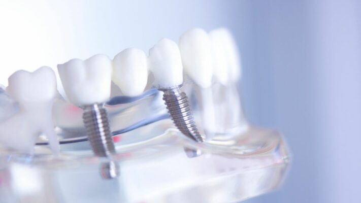
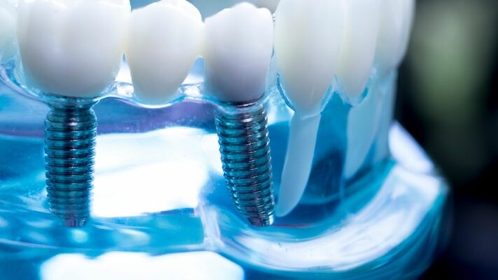
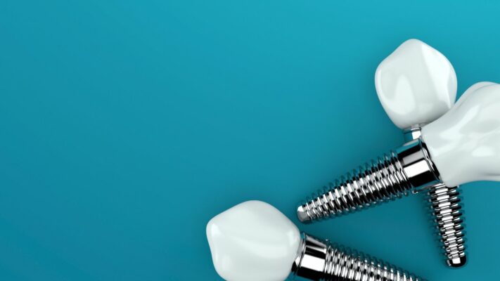
What are modern approaches in tooth extraction and implant treatments?
Oral surgery and implantology focus on replacing lost teeth and preserving existing bone tissue during this process.
What can be done to prevent bone loss after tooth extraction?
Tooth extraction inevitably initiates a healing and remodeling process in the bone. Unfortunately, this process always results in bone resorption (loss). Within the first 6 months after extraction, a significant loss in both the width and height of the bone may occur.
There are two modern approaches to minimize this loss. The first is “minimally traumatic extraction.” The goal of this technique is to remove the tooth without breaking or damaging the thin bone wall, especially on the cheek (buccal) side. This thin wall is the most prone to resorption.
The second is the “Socket Preservation” (Alveolar Ridge Preservation – ARP) procedure. This is performed to preserve the extraction socket and is particularly indicated in cases where the implant will not be placed immediately (delayed implant). After minimally traumatic extraction, the socket is thoroughly cleaned of infected tissues, and a bone graft (bone substitute) material is placed inside the socket. It is usually covered with a resorbable collagen membrane.
This procedure cannot completely stop bone loss; it can only limit it. Its main clinical benefit is that it prepares an ideal foundation for future implant placement by minimizing the need for more complex, costly, and challenging bone augmentation procedures (bone regeneration or sinus lifting).
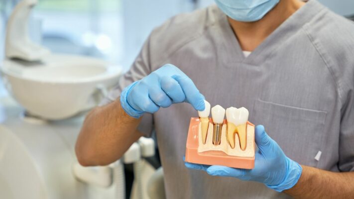
Can an implant be placed immediately after tooth extraction?
The timing of implant placement after tooth extraction is one of the most critical decisions in treatment. There are three main timings:
- Immediate
- Early (4–16 weeks later)
- Delayed (4 months and later)
“Immediate implant,” meaning placing the implant in the same session as tooth extraction, is possible and advantageous when certain conditions are met. However, this is a complex topic that requires careful interpretation of clinical data.
Scientific studies tend to show that in terms of implant survival (success) rates, delayed implants (placed after waiting) are statistically slightly safer and more predictable than immediate implants. In delayed implants, success rates are consistently above 95%.
The main difference emerges in complications and aesthetics. This is a “high risk, high reward” scenario:
Risk: Immediate implant placement is associated with a higher risk of complications. The most prominent risk is gum recession in the area where the implant is placed.
Reward: On the other hand, some evidence suggests that in ideal cases (for example, when the extraction socket is completely intact and the patient has a thick gum type), the final aesthetic outcome may be better with immediate implants.
This shows that case selection is paramount. While delayed implant placement appears to be a safer, lower-risk, and more predictable “safe harbor” protocol, immediate implant placement may offer better aesthetic results when ideal conditions are met.
In single-tooth loss, is a bridge or an implant better?
In the case of a single missing tooth, an implant-supported single crown (ISSC) is clinically superior to a traditional tooth-supported bridge (FDP).
Implants are much more conservative and less invasive because they do not require cutting (reducing) the adjacent (often healthy) teeth next to the missing tooth, which is necessary for a bridge. Only the missing area is treated.
In addition, implant-supported crowns are more long-lasting. Over a 5-year follow-up, the survival rate of implant-supported crowns is 96.8%, whereas this rate is 84% for traditional bridges. When a problem occurs with a bridge (for example, if one of the supporting teeth decays), the entire structure must be replaced, whereas an implant is a single independent structure.
Are clear aligners or braces more effective in orthodontic treatment?
Orthodontics corrects crowding and bite problems in the teeth and jaws. Today, two main methods stand out: clear aligners and traditional fixed appliances (braces).
Scientific reviews confirm that both methods are generally effective in straightening teeth. When compared, distinct advantages and disadvantages emerge.
Evidence shows that the total treatment duration of clear aligner treatments is significantly shorter than that of fixed braces. In addition, aligners offer advantages such as aesthetics, comfort, and removability.
However, although overall success scores are similar, fixed braces are still superior to clear aligners in achieving certain specific and challenging details.
Braces are more effective than clear aligners in the following areas.
- Precisely controlling tooth “torque” (root position)
- Completing difficult rotations, especially in round teeth such as premolars
- Ensuring proper occlusal contact (bite fit) at the end of treatment
This shows that clear aligners are an excellent tool for correcting mild to moderate crowding, but very challenging cases requiring significant root movement or precise bite adjustment are still treated more predictably with braces.
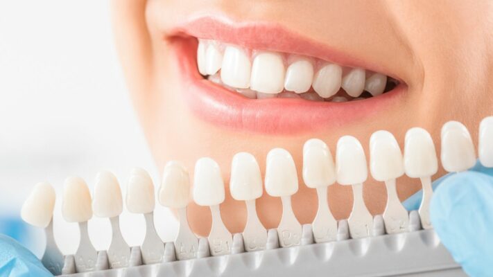
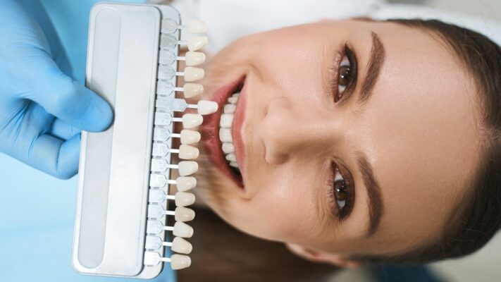
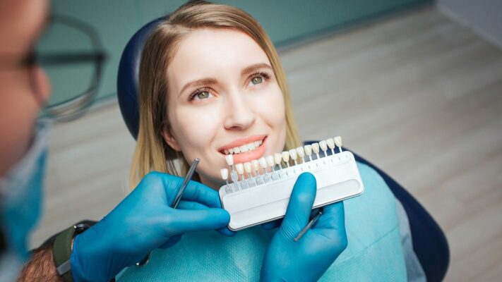
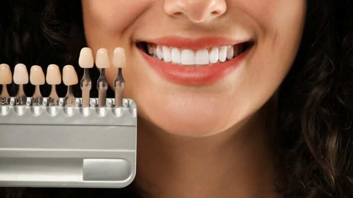
What are tooth whitening and aesthetic laminate treatments?
Aesthetic dentistry includes tooth whitening (bleaching) and veneer (laminate) treatments that focus on improving the color and shape of teeth.
Can a root canal-treated tooth be whitened if it has changed color?
Yes. This procedure is called “non-vital bleaching” or “internal bleaching” (walking bleach). It is applied only to teeth that have undergone root canal treatment and have changed color internally over time (turned gray or brown).
In the procedure, the old filling at the back of the tooth is accessed, and the top of the root canal filling is opened. To protect the root, a protective “barrier” filling is обязательно placed over the root canal filling. Then a whitening agent is placed inside the tooth cavity, and it is sealed with a temporary filling. The color is checked after a few days, and once the desired shade is achieved, the procedure is completed. The most critical and mandatory step in this procedure is that barrier filling placed to protect the root.
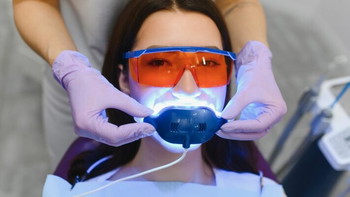
Is in-office whitening or at-home whitening more long-lasting?
For vital teeth, there are two main professional methods: “In-Office” whitening (fast results with high-concentration agents performed in the clinic) and “At-Home” whitening (nightly use of low-concentration agents with custom trays under dentist supervision).
Effectiveness: Numerous scientific reviews have concluded that there is no significant difference between the two methods in terms of final color change (whitening). Both effectively whiten teeth.
Speed and Longevity: This is the most important difference. In-office whitening provides very fast results (1 hour). At-home whitening is slower (takes 1–2 weeks). However, in terms of longevity, evidence suggests that at-home whitening may provide longer-lasting whiteness compared to in-office whitening and that color relapse may be less likely.
The clinical decision is related not to effectiveness but to patient compliance and longevity management. In-office whitening is faster but color may return sooner. At-home whitening is slower and requires high patient compliance, but results may be more durable.
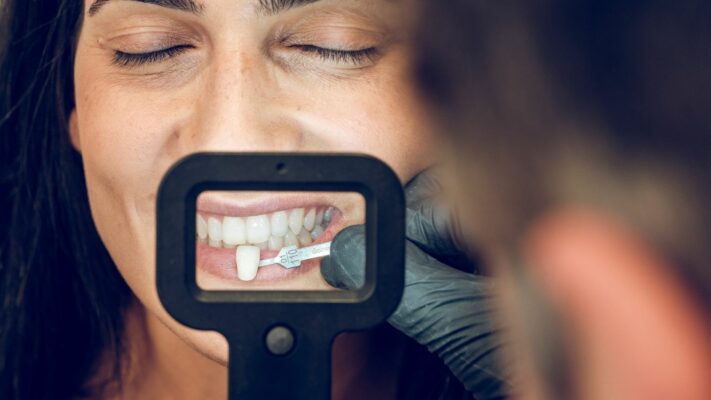
What is the difference between porcelain veneers and composite veneers (bonding)?
Veneers (laminates) are thin layers bonded to the front surface of teeth. They are used to correct permanent discolorations, fractures, wear, gaps between teeth, or minor shape irregularities. There are very clear differences between the two main materials in terms of durability, aesthetics, and lifespan:
Porcelain (Ceramic) Veneers:
- Lifespan: 10–20 years (long-term)
- High resistance to staining (tea, coffee, smoking do not affect color)
- Superior and long-lasting aesthetics
- Produced in a laboratory (higher cost)
- Higher fracture resistance
Composite Veneers (Bonding):
- Lifespan: 5–7 years (medium-term)
- Risk of staining and color change over time
- Good aesthetics (but not as bright and natural as porcelain)
- Can be applied in a single session in the clinic (more affordable)
- Less resistant to wear and fracture (easy to repair)
The difference in lifespan between the two treatments (10–20 years for porcelain, 5–7 years for composite) actually places them in different categories. Porcelain veneers are long-term prosthetic restorations and, considering their total lifespan, are generally a more permanent solution. Composite veneers, on the other hand, serve as a more conservative (may require less tooth reduction), faster, and lower initial cost alternative.
Blog Posts

Most Preferred Countries for Hair Transplantation
Among the most preferred countries for hair transplantation are Turkey, India, South Korea, and Poland. [...]
Read More
21
Jan
Jan

Hair Transplant Prices
Hair transplant prices vary depending on the technique to be used, the number of grafts, [...]
Read More
21
Jan
Jan
Most Preferred Countries for Plastic Surgery
Among the most preferred countries for plastic surgery, Turkey, South Korea, the United States, and [...]
Read More
21
Jan
Jan

Plastic Surgery Prices
Plastic surgery prices vary depending on the type of procedure, geographical location, the surgeon’s experience, [...]
Read More
21
Jan
Jan

Physical Therapy Prices
Physical therapy prices vary depending on the number of sessions to be applied, the treatment [...]
Read More
21
Jan
Jan

Dental Treatment Prices
Dental treatment prices vary depending on the type of procedure to be performed, the quality [...]
Read More
21
Jan
Jan
Most Preferred Countries for Dental Treatments
Among the most preferred countries for dental treatments are Turkey, Hungary, Mexico, and Thailand. These [...]
Read More
21
Jan
Jan