
 Türkçe
 Deutsch
 Français
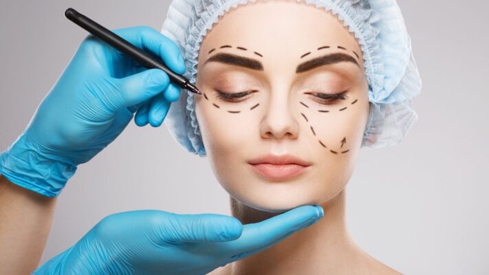
Eyelid surgery, medically known as blepharoplasty, is a procedure in which the anatomical structures around the eyes are surgically rearranged. This procedure includes interventions that directly affect the contour and structure of both the upper and lower eyelids. Surgical techniques aim to reshape the area by focusing on the main components of the eyelid. Blepharoplasty is defined as a specific surgical approach, planned according to the current condition of the eyelids, that changes the appearance of the area.
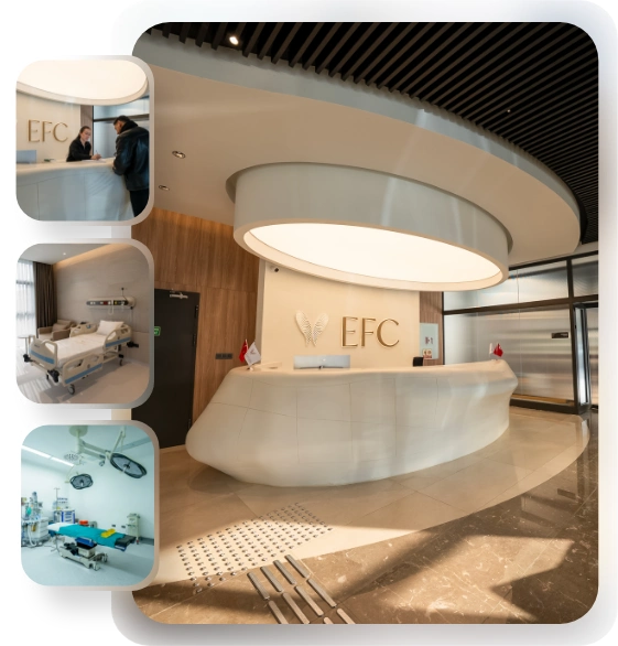
EFC CLINIC
Comprehensive Care: From Initial Consultation to Follow-Up.
EFC CLINIC is a center of excellence specializing in the most meticulous fields of surgical medicine, from aesthetic surgery to interventional treatments—where every step progresses with refined attention. Medical excellence, aesthetic precision, and uncompromising ethical standards converge on the same path. Our subspecialty-trained experts aim to achieve natural and reliable results by delivering evidence-based care supported by modern imaging, standardized protocols, and safety systems. From consultation to recovery, your care is coordinated end-to-end with clear communication, transparent planning, and genuine respect for your health.
Why is a doctor’s examination so important for a successful eyelid surgery?
The success of eyelid surgery depends not only on the technique used but perhaps even more on the correct diagnosis made before surgery. Patients usually come with general complaints such as “my eyes look very tired.” However, there can be many different underlying reasons for this ‘tired’ appearance.
The most common cause of an insufficient or unsatisfactory surgical result is performing the right surgery for the wrong diagnosis. For example, if a patient’s main problem is drooping eyebrows (brow ptosis), failing to notice the skin that this condition piles onto the eyelid and removing only the eyelid skin does not solve the underlying problem. It can even cause the eyebrows to droop further and distort the expression. Therefore, clearly identifying the source of the problem at the first examination is more important than the surgery itself.
What different anatomical problems can underlie the ‘tired’ appearance of the eyelid?
There is not a single cause for that ‘tired’ look around the eyes. During the examination, it is necessary to clearly identify the actual anatomical problem or combination of problems causing this appearance. These problems may include:
- Dermatochalasis (True excess skin)
- Steatoblepharon (Fat herniation/bagging)
- Blepharoptosis (Drooping of the eyelid itself)
- Brow ptosis (Drooping of the eyebrow)
It is important to understand the differences among these problems. ‘Dermatochalasis’ is the actual loosening and sagging of the eyelid skin; this is the most common cause we see. ‘Steatoblepharon’ is the forward herniation of fat as a result of weakening of the membrane (orbital septum) that holds the fat pads surrounding the eye in place. This condition causes the bulging appearance known as ‘bags,’ especially in the lower eyelids.
‘Blepharoptosis’ (often abbreviated as ptosis) is a completely different condition. It is not an excess skin problem, but the eyelid margin (where the eyelashes emerge) sits lower than normal due to weakening of or separation of the muscle that elevates the eyelid (the levator muscle). This makes the eye look ‘droopy’ or ‘half-closed.’
‘Brow ptosis’ is the downward descent of the eyebrows with age, especially from the outer parts, due to loss of volume. This drooping pushes the upper eyelid skin downward, causing it to bunch up and create a ‘pseudo’ excess skin appearance. Very often, a significant number of patients have more than one of these problems together:
When is eyelid aesthetics a medical necessity, and when is it considered a cosmetic preference?
Eyelid surgery (blepharoplasty) can be performed for both functional (i.e., a medical necessity) and cosmetic (i.e., aesthetic) reasons.
What Are the Medical Necessity (Functional) Reasons?
In these cases, the anatomical abnormality of the eyelid objectively affects the person’s quality of life or health. The main conditions in which the procedure is considered medically necessary are as follows:
- Restriction of the visual field
- Chronic eye fatigue (asthenopia)
- Compensatory forehead headaches
- Need to look by constantly keeping the chin raised
- Rash and irritation between skin folds (contact dermatitis)
- Eyelashes rubbing against the eye or turning inward
At the top of these is restriction of the visual field. Sagging upper eyelid skin or a drooping eyelid margin can descend over the eyelashes and physically obstruct a person’s vision, especially upward and to the sides. This can be objectively documented with a visual field test called ‘perimetry.’ In addition, patients often have to chronically contract the ‘frontalis’ muscle in the forehead to lift these drooping lids and see better. This constant effort can lead to significant eye fatigue and chronic headaches.
What Are the Cosmetic (Aesthetic) Reasons?
In this case, the procedure is chosen to improve the person’s appearance rather than due to medical necessity. Patients’ complaints on this topic are usually as follows:
- A tired, sleepy, or exhausted appearance
- Taking on a sad or angry expression
- More prominent under-eye bags
- An overall aged appearance
- A desire to have a more vibrant, rested, and youthful look
Cosmetic blepharoplasty helps restore overall facial harmony and soften the tired expression caused by aging.
Which critical points are examined during an eyelid evaluation?
A complete evaluation is essential to choose the correct surgical technique and, most importantly, to avoid complications.
Contact us now to get detailed information about our treatments and procedures and to schedule an appointment!
Why Is Eye Health History Important?
The first step in avoiding complications is listening to the patient’s history. In particular, symptoms related to ‘Dry Eye Syndrome’ (DES) (burning, stinging, tearing) must be questioned. Because temporary swelling after surgery and incomplete eyelid closure can significantly worsen an underlying dry eye condition. Those who have previously undergone laser vision correction such as LASIK or those receiving certain hormonal treatments may be at higher risk in this regard, and some precautions may be needed before surgery.
What Is Evaluated in the Physical Examination?
The periocular area is a whole; the eyelid, eyebrow, and cheek are anatomically connected. In the examination, all these structures are assessed as a whole. The main evaluation points are as follows:
- Eyebrow position and mobility
- Level of the upper eyelid margin (Ptosis check)
- Lower eyelid laxity (Laxity test)
- Cheek–globe relationship (Vector analysis)
- Festoons (Malar bags) and the tear trough
The assessment must start with the eyebrow position. The doctor lifting the brow by hand during the examination allows them to distinguish ‘true’ excess skin from the ‘pseudo’ excess caused by brow drooping. By looking at where the upper lid margin (the base of the eyelashes) sits relative to the pupil, it can be determined whether there is ‘ptosis’ (drooping of the eyelid).
For the lower lid, laxity is checked with the ‘snap test’ (pull-and-release test). Because a lax lid carries a risk of postoperative downward pull or outward turning (ectropion). In addition, the adequacy of cheek support (negative vector) is assessed, and it is checked whether there are edematous pouches called ‘festoons’ (malar bags) located over the cheekbone, which are different from under-eye bags.
How is a standard upper eyelid surgery (Blepharoplasty) performed?
Upper eyelid surgery, i.e., standard blepharoplasty, is basically performed to eliminate excess skin (dermatochalasis) and, if present, fat bagging (steatoblepharon).
During surgery, an incision is made along the natural eyelid crease line. Since this incision is placed exactly in the fold, the scar becomes almost invisible once healed. The amount of skin to be removed is measured very precisely so that it will not prevent the eye from closing fully. Usually, together with this piece of skin, a thin underlying strip of loosened muscle (the orbicularis muscle) is also removed. Then the membrane called the ‘orbital septum,’ which holds the fats in place, is opened to access the herniated fat pads.
There are two main fat pads (central and medial/nasal) in the upper eyelid. Laterally, there is the lacrimal gland; it is very important to distinguish this gland from fat and to preserve it, because accidental injury can cause chronic dry eye.
The surgical philosophy in fat management has changed greatly over the years. In the past, with an aggressive “debulking”-focused approach, as much fat and tissue as possible would be removed. However, over time this caused a “hollow,” “sunken,” and “skeletonized” periocular appearance. Today’s modern approach is strictly “volume-preserving.” That is, only the truly herniated excess fats are removed or contoured in a very conservative way. The goal is to provide a fresher and more rested appearance without losing the natural fullness around the eyes.
How are eyelid drooping (Ptosis) and excess skin corrected at the same time?
In a significant number of patients, there is not only excess skin but also drooping (ptosis) of the eyelid margin itself. If these two problems coexist, it is possible—and the most correct approach—to solve both with a single surgery.
In this combined approach, the standard eyelid aesthetic (blepharoplasty) incision is again used. Through this incision, the surgeon first reaches the deeper underlying problem, namely the weakened or detached tendon (aponeurosis) of the muscle that elevates the eyelid (levator).
This deep repair is performed first. The muscle tendon is found, strengthened, or reattached with sutures to where it should be, namely the cartilage structure of the lid (tarsus). If the procedure is performed under local anesthesia, the patient may be asked to open and close the eye at this stage so that the symmetry of lid height and curvature can be adjusted instantly on the operating table.
After the eyelid margin height is brought to its ideal position, the procedure returns to the superficial plane. The excess skin and muscle tissue planned at the beginning are removed, and the blepharoplasty is completed. In other words, through a single incision, both the eyelid-lifting muscle is repaired (ptosis correction) and the excess skin is removed (blepharoplasty).
Why is Asian eyelid surgery (‘double eyelid surgery’) different?
Asian eyelid surgery is anatomically completely different from Western blepharoplasty. This is because about half of individuals of Asian origin have a ‘single eyelid’ (monolid), meaning there is no upper eyelid crease line (supratarsal crease).
The main anatomical reasons for this are as follows:
- The tendon of the eyelid-lifting muscle (levator) does not attach strongly enough to the skin
- The muscle tendon attaches to the eyelid margin at a much lower level
- As a result, the periocular fat pads sag lower, creating a ‘puffy’ appearance
- A prominent ‘epicanthal fold’ at the inner corner of the eye
The goal of this surgery is not to “Westernize” or create a very high, arched lid. On the contrary, this is generally an undesired and unnatural-looking result. The aim is to create a natural-looking ‘double eyelid’ crease that makes the eye appear larger and more vivid while preserving the patient’s ethnic features. This is done using special suture techniques or by creating a muscle–skin connection through an incision to establish a new fold location in the skin. Sometimes ‘epicanthoplasty’ may also be added to open the fold at the inner corner.
Which surgical approaches are available for lower eyelid bags?
Lower eyelid rejuvenation is anatomically more complex than the upper eyelid and requires very delicate handling of the lid–cheek junction. Basically, there are two different surgical approaches. The choice of technique depends entirely on whether the patient has excess skin on the lower eyelid.
Contact us now to get detailed information about our treatments and procedures and to schedule an appointment!
When is lower eyelid surgery performed through an ‘under-lash’ incision (Transcutaneous)?
This approach is necessary for patients who have not only fat bagging in the lower eyelid but also significant excess skin and muscle laxity (dermatochalasis). In short, this method is chosen if some skin must be removed and the loosened muscle needs to be tightened.
The incision is made 1–2 mm just below the eyelashes, along a line called the ‘subciliary’ line. This scar is usually hidden in a natural crease. Entering through this line, the skin and the underlying muscle tissue (skin–muscle flap) are elevated as a single unit. This gives the surgeon full access to all three fat pads underneath. After the fat bags are managed (removed or repositioned), the skin–muscle flap is gently laid back into place.
The most critical point of this method is removing the skin very conservatively (in a very small amount). Aggressively removing too much skin to make it “tighter” is the most important cause of downward pull (retraction) of the lower eyelid or outward turning (ectropion), the most feared complication. Therefore, the surgeon’s experience is vital at this point.
Who is suitable for scarless lower eyelid aesthetics (Transconjunctival)?
This is the ideal and preferred method especially for patients (generally younger) who have no excess skin and complain only of under-eye bags (fat herniation).
In this technique, no incision is made on the outside skin. The incision is made from the inner surface of the lower eyelid (the pink, moist tissue called the conjunctiva). This is like a ‘hidden’ route and provides direct access to the posterior surface of the fat pads. The surgeon can remove the herniated fats through this internal incision or reposition them (fat transposition).
The biggest advantages of this method are as follows:
- There is no visible external scar.
- The healing process is usually faster.
- Most importantly, it is much safer for the position of the lower eyelid.
Because the skin, muscle, and the orbital septum membrane that holds them in place are not cut in this technique (i.e., the anterior and middle support structures of the lid are not violated), the risk of downward pull or outward turning (ectropion) that can be seen with an under-lash incision is almost nonexistent with this technique. If skin removal is not required, this method is always the superior choice.
What should be done with the fat in under-eye bags: Should the fat be removed, or should it be repositioned?
In lower lid surgeries, fat management is perhaps the most important step that determines the naturalness of the result. In the past, these fats were seen as “excess” and were removed as much as possible. However, we now know that this causes the under-eye area to become “sunken” and “hollow,” creating an older and more tired appearance.
- Fat Removal (Excision)
This is simply removing the fat bag. It is now preferred only for a very small group of patients (usually young patients) who do not have a hollow called the ‘tear trough’ and have only a prominent fat bag. Removing fat from a patient who has an under-eye hollow can worsen that hollow.
- Fat Repositioning (Transposition / Redraping)
This is today’s modern and standard approach. It is especially applied in patients who have both an under-eye bag (swelling) and a tear trough (hollow).
In this technique, the herniated fat that causes the ‘bag’ appearance is not removed. On the contrary, this valuable tissue is preserved. The fat pad is released and, from where it is bulging, it is spread like a quilt into the hollow area just below called the ‘tear trough’ (redrape/reposition). This fat is fixed either under or over the periosteum.
The purpose of this method is to use the patient’s own fat as a natural “filler.” In this way, both the swelling (bag) is eliminated and the hollow (trough) is filled. This provides a smooth, youthful, and convex transition at the lid–cheek junction.
Why is a ‘suspension’ (Canthopexy) procedure necessary in lower eyelid surgery?
Lower eyelid surgeries, especially when an incision is made under the lashes, carry a risk for the natural position and tightness of the lid. If the patient’s lid was already found to be lax in the preoperative examination, this risk is much higher. Procedures such as ‘canthopexy’ or ‘canthoplasty,’ although sometimes used to create an “almond eye” aesthetic, are primarily critical support procedures that reduce this risk.
In the preoperative examination, lid laxity must be tested with the ‘snap test’ (pull-and-release). If there is laxity, a support procedure must be performed in the same session at the outer corner of the lid (lateral canthal tendon) to prevent outward turning (ectropion) or sagging.
- Canthopexy: A less invasive procedure. It is the mild tightening of the lateral canthal tendon by reinforcing it with sutures and suspending it to the periosteum. It is generally done for mild laxity or for preventive purposes.
- Canthoplasty: A more comprehensive repair. The canthal tendon is released, shortened if necessary, and re-suspended more firmly to the lateral orbital rim bone (anchoring). It is required in cases of severe laxity or existing lid sagging.
How does a brow lift affect the outcome of eyelid aesthetics?
The eyelid and the brow cannot be considered separately; they are a single aesthetic unit. Sometimes patients come with the complaint “my eyelids are very saggy,” but on examination it is seen that the main issue is that their brows, especially the outer part, have drooped downward due to gravity (brow ptosis).
A drooping brow piles the eyelid skin onto itself and creates a ‘pseudo’ excess skin (pseudo-dermatochalasis). If this diagnosis is missed and skin is aggressively removed only from the eyelid, the result is unsuccessful. The brow is pulled even further down and gives the person an ‘angry’ or more tired expression.
The correct approach is first to correct the underlying problem, the brow droop (for example with an endoscopic, temporal, or direct brow lift). After the brow is brought to its ideal position, it is assessed how much true excess skin remains on the eyelid. Most of the time, after a brow lift, either very little skin removal is needed or sometimes none is necessary. Combining the two procedures when indicated provides a much more harmonious, natural, and comprehensive rejuvenation of the upper face.
How are botulinum toxin (Botox) injections used for wrinkles around the eyes?
Periocular rejuvenation is not only surgical. Eyelid surgery corrects structural problems (sagging skin and herniated fat), but it does not by itself correct skin quality or expression wrinkles.
Botulinum toxin (BoNT) injections are used for ‘dynamic’ wrinkles, meaning those that occur with movement (smiling, frowning). By temporarily blocking signal transmission at the nerve endings of the relevant muscles, it allows the muscle to relax and the skin over it to become smoother.
The main areas of use around the eyes are as follows:
- Lateral canthal lines (Crow’s feet)
- Glabellar lines (Frown lines or ’11’ lines)
- Forehead lines
- Chemical brow lift (a subtle application that lifts the lateral tail of the brow)
How are under-eye hollows treated with tear trough filler?
Hyaluronic Acid (HA)-based fillers are used to non-surgically correct static hollows and volume loss around the eyes. The most common area of application is the ‘tear trough’ deformity. It can also be considered a non-surgical alternative to fat transposition surgery performed for under-eye bags, but its effect is temporary.
Filler in this area is a procedure that requires very high technical precision.
Things to Consider in Under-Eye Filler:
- Correct Filler Selection: Because the under-eye skin is very thin, fillers produced specifically for this area, with fewer cross-links and low water-retention capacity (low hydrophilic), should be preferred. Otherwise, excessive swelling or the ‘Tyndall effect’ (a bluish discoloration visible under the skin) may be seen.
- Correct Injection Plan: The filler should not be placed just under the skin, i.e., superficially. This leads to visible lumps and irregularities. The injection should be performed under the muscle, directly on the bone (supraperiosteal), i.e., in the deepest plane.
- Correct Volume: The under-eye area does not tolerate “overcorrection.” Applying a small amount (under-correction) and, if necessary, performing a touch-up after 2–3 weeks always provides a safer and more natural result.
Results are temporary, usually lasting 6–12 months, but when performed correctly, patient satisfaction in reducing a tired expression is very high.
What is the recovery process after eyelid surgery, and what are the possible complications?
The postoperative recovery process is generally fast and comfortable.
The first 1–2 weeks are the acute period with swelling and bruising. Vision may be temporarily blurred due to antibiotic ointments used or edema. Sutures are usually removed on days 5–7. Patients can generally return to social life and wear makeup within 7–10 days. It may take a few months for the swelling to fully subside and for the final result to settle. During this period, using sunscreen is very important to keep scars from being exposed to the sun.
Although blepharoplasty is generally a safe procedure, like any surgery it carries risks and complications. The most important complications are as follows:
- Temporary dry eye (DES)
- Lagophthalmos (Inability of the upper lid to close completely)
- Ectropion (Outward turning of the lower lid)
- Retraction (Downward pull of the lower lid)
- Retrobulbar hematoma (Bleeding behind the eye)
Dry Eye: It is the most common side effect after surgery and is usually temporary. It results from the ocular surface being slightly exposed due to swelling and is easily managed with artificial tears.
Lagophthalmos: This is the inability of the eye to close completely due to excessive skin removal from the upper lid. It is a serious complication, and measurements must be made very carefully to prevent it.
Ectropion and Retraction: This is the most feared complication of lower lid surgery. It usually results either from excessive skin removal or from failure to identify and support lid laxity beforehand (not performing canthopexy). Most of these complications are preventable with correct surgical planning and a conservative approach. If it occurs, it is initially observed with massage and taping; if it does not resolve within 6 months, surgical repair is required.
Retrobulbar Hematoma: This is a very rare but vision-threatening emergency. Blood collects behind the eye after surgery, compresses the optic nerve, and requires urgent surgical intervention (decompression).
Blog Posts
Most Preferred Countries for Hair Transplantation
Among the most preferred countries for hair transplantation are Turkey, India, South Korea, and Poland. [...]
Read More
21
Jan
Jan
Hair Transplant Prices
Hair transplant prices vary depending on the technique to be used, the number of grafts, [...]
Read More
21
Jan
Jan
Most Preferred Countries for Plastic Surgery
Among the most preferred countries for plastic surgery, Turkey, South Korea, the United States, and [...]
Read More
21
Jan
Jan
Plastic Surgery Prices
Plastic surgery prices vary depending on the type of procedure, geographical location, the surgeon’s experience, [...]
Read More
21
Jan
Jan
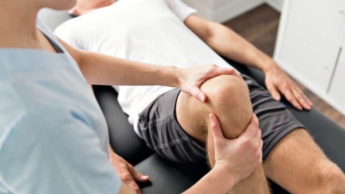
Physical Therapy Prices
Physical therapy prices vary depending on the number of sessions to be applied, the treatment [...]
Read More
21
Jan
Jan

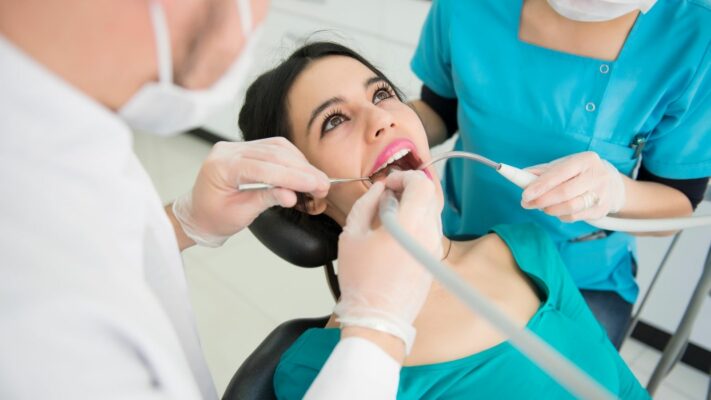
Dental Treatment Prices
Dental treatment prices vary depending on the type of procedure to be performed, the quality [...]
Read More
21
Jan
Jan
Most Preferred Countries for Dental Treatments
Among the most preferred countries for dental treatments are Turkey, Hungary, Mexico, and Thailand. These [...]
Read More
21
Jan
Jan